Cilia
and flagella are tiny waving filaments with important functions in humans, such
as clearing our airways. A 3D electron microscopy study of the normal (WT) and
mutant varieties of flagella has revealed new details of the structure, protein
composition, and connections between neighboring components formed by a crucial
structural complex of this biological nanomachine,
the "nexin-dynein
regulatory complex" (N-DRC), which is shown in color on the left. In the poorly
moving mutants, this structure is damaged or almost missing. This opens the way
for building a new mechanical model of this device, and how it functions, along
with mechanical testing of individual cilia and flagella, normal and mutant, in
the Brandeis multi-mode optical microscopy laboratory.
Cilia and flagella
are highly conserved sensing and motility organelles of eukaryotes and ciliary defects have been
linked to many human diseases. The core structure of cilia and flagella, the axoneme, consists of a
central microtubule pair and nine surrounding microtubule doublets that are
connected by nexin links. Movement of
cilia and flagella is generated by precisely orchestrated activity of many
thousands of dynein motors. One of the
key regulators of this activity is the dynein
regulatory complex (DRC), but detailed structural information has been missing.
By comparing the DRC structure of wild-type, 4 different drc-mutants and 1 drc-mutant rescue we
were able to visualize the DRC in situ at molecular resolution, and to
deconstruct a key complex in this biological nanomachine. This work has now been published (Heuser et
al. 2009, JCB 187:921)
Recently, the PIs Dogic, Fraden and Nicastro obtained outside
funding from the W.M. Keck Foundation for studying "Active Matter", including
the present seed project.
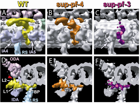
These images show
lateral- and end-views of the basic structural element of the flagellum,
including the long hollow microtubules and associated proteins, like motors and
regulators. Normal structure of the N-DRC, a regulatory bridge (colored), at
the left (WT), a mild mutant of the complex in the middle (sup-pf-4) and a
mutant with more severe defects on the right (sup-pf-3). These images were
obtained by cryo-electron tomography,
revealing the 3D structure at the molecular scale.